Leg Bone Diagram : Human Skeleton Labeled Diagram . Human Skeleton Labeled ... / The answer is still unknown, but hereditary.
Leg Bone Diagram : Human Skeleton Labeled Diagram . Human Skeleton Labeled ... / The answer is still unknown, but hereditary.. The largest and most medial leg bone, forming both the knee and ankle joints. Bone basics and bone anatomyhave you ever seen fossil remains of dinosaur and ancient human bones in textbooks, television, or in person at a museum? This framework consists of many individual bones and cartilages. The axial skeleton and the appendicular formed by the left and right hip bones, the pelvic girdle connects the lower limb (leg) bones to the axial. Human skeleton, the internal skeleton that serves as a framework for the body.
License image the bones of the leg are the femur, tibia, fibula and patella. This framework consists of many individual bones and cartilages. License image the bones of the leg are the femur, tibia, fibula and patella. Pelvis definition, anatomy, diagram, & facts. These bones are arranged into two major divisions:

They allow you to move and provide support for your upper body.
However, the definition in human anatomy refers only to the section of the lower limb extending from the knee to. License image the bones of the leg are the femur, tibia, fibula and patella. Skeleton leg ankle joints and toe phalanges, cuboid, metatarsal, navicular and cuneiform bones, hand drawn dorsal view of foot. Ankle and foot pain massage therapy foot treatment. These bones have a marrow, but not a bone marrow cavity. Each leg is made up of four bones. Your leg bones are the longest and strongest bones in your body. The foot bones shown in this diagram are the talus, navicular, cuneiform, cuboid, metatarsals and calcaneus. Click now to learn more about the bones, muscles, and soft tissues tibia: These bones are arranged into two major divisions: However, the definition in human anatomy refers only to the section of the lower limb extending from the knee to the ankle, also known as the crus or. Quizzes on human skeletal system anatomy, bone anatomy, and bone markings. Normal leg bones are relatively straight, but those affected by paget's disease are porous and curved.
Time to jump right into the biggest and strongest bones in the human body. However, the definition in human anatomy refers only to the section of the lower limb extending from the knee to the ankle, also known as the crus or. The foot bones shown in this diagram are the talus, navicular, cuneiform, cuboid, metatarsals and calcaneus. The answer is still unknown, but hereditary. However, the definition in human anatomy refers only to the section of the lower limb extending from the knee to.
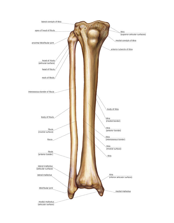
The bones of the leg are the femur, tibia, fibula and patella.
There also are bands of fibrous connective tissue—the. The axial skeleton and the appendicular formed by the left and right hip bones, the pelvic girdle connects the lower limb (leg) bones to the axial. Ankle and foot pain massage therapy foot treatment. Pelvis definition, anatomy, diagram, & facts. The foot bones shown in this diagram are the talus, navicular, cuneiform, cuboid, metatarsals. Each leg is made up of four bones. You'll learn about the muscles, bones, and other structures of each area of the leg. Skeleton leg ankle joints and toe phalanges, cuboid, metatarsal, navicular and cuneiform bones, hand drawn dorsal view of foot. Click now to learn more about the bones, muscles, and soft tissues tibia: Learn how to draw the femur, patella, tibia, and fibula in this lesson! The foot bones shown in this diagram are the talus, navicular, cuneiform, cuboid, metatarsals. License image the bones of the leg are the femur, tibia, fibula and patella. Quizzes on human skeletal system anatomy, bone anatomy, and bone markings.
Muscles that lift the arches of the feet. Human skeleton, the internal skeleton that serves as a framework for the body. The axial skeleton and the appendicular formed by the left and right hip bones, the pelvic girdle connects the lower limb (leg) bones to the axial. Ankle and foot pain massage therapy foot treatment. What causes the osteoclasts to become overactive?

However, the definition in human anatomy refers only to the section of the lower limb extending from the knee to.
These bones are arranged into two major divisions: Human bone diagram wiring diagrams click. Bones of the leg and foot, lower leg bone anatomy, leg bones anatomy, leg muscles, leg bones diagram, leg bone structure, leg anatomy muscles, parts of the lower leg. Normal leg bones are relatively straight, but those affected by paget's disease are porous and curved. Most relevant best selling latest uploads. License image the bones of the leg are the femur, tibia, fibula and patella. Learn vocabulary, terms and more with flashcards, games and other study tools. Muscles that lift the arches of the feet. Time to jump right into the biggest and strongest bones in the human body. This framework consists of many individual bones and cartilages. In the leg, the interosseous membrane extends between the tibia and the fibula, running along the crests of the bones. It's easy to look at these and think of. Diagram of human bone anatomy.


Komentar
Posting Komentar